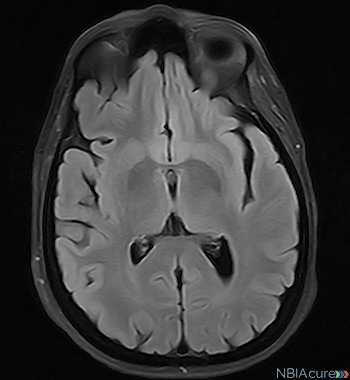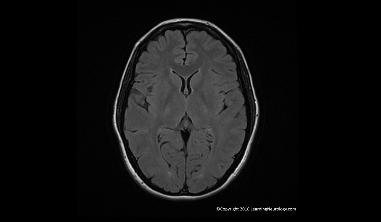

They nullify certain tissue types based on their inversion timings, thereby stopping tissues such as fat and CSF from appearing as bright signals.

These types of images are manipulations of T1 and T2. Meningioma is shown more clearly by gadolinium contrast with a dural tail 5 Meningiomas will have a homogenous enhancement after the contrast, but will also have a “dural tail,” meaning the lesion appears continuous with the dura (Figure 2). Typical intracranial abscesses have a “ring-enhancement” pattern, while metastases enhance homogeneously. Gadolinium appears bright in signal, allowing for detection of detailed abnormalities (e.g.

This process involves injecting 5-15ml of contrast intravenously, with images taken shortly thereafter. arteries) or pathologically-vascular tissues (e.g. This is useful in adrenal tumours or bone marrow pathology, where the image will appear homogenous with surrounding tissue due to fat content. The fat signal can be suppressed to enable a better view of pathology in and around anatomical structures – particularly oedema. Normal brain MR shows differences between T1 and T2 images 3 Additional features of T1/T2 weighted images

1 Consideration should be given to patients who are claustrophobic as well. MRI is contraindicated in patients who have ferromagnetic metal implants or foreign bodies. It takes slightly longer to acquire MR images and they are more expensive. MRI is particularly helpful in patients with suspected neurological or musculoskeletal pathology, however, they can be used in many other specialities too. They are often reserved for superior viewing of soft tissues. Generally, MRI is used less commonly than plain films and CT scans. You might also be interested in our OSCE Flashcard Collection which contains over 2000 flashcards that cover clinical examination, procedures, communication skills and data interpretation.


 0 kommentar(er)
